 Get the Genesis
of Eden AV-CD by secure
internet order >> CLICK_HERE
Get the Genesis
of Eden AV-CD by secure
internet order >> CLICK_HERE
Windows / Mac Compatible. Includes live video seminars, enchanting renewal songs and a thousand page illustrated codex.
Get the Genesis
of Eden AV-CD by secure
internet order >> CLICK_HERE
Windows / Mac Compatible. Includes
live video seminars, enchanting renewal songs and a thousand page
illustrated codex.
 Return
to Genesis of Eden?
Return
to Genesis of Eden?
The Three-pound Universe
For good reason, the human brain is sometimes hailed as the most complex object in the universe. It comprises a trillion cells, 100 billion of them neurons linked in networks that give rise to intelligence, creativity, emotion, consciousness and memory. Large anatomic subdivisions in the brain offer a rough map of its capabilities. At a very gross level, the brain is bilaterally symmetric, its left and right hemispheres connected by the corpus callosum and other axonal bridges. Its base consists of structures such as the medulla, which regulates the autonomic functions (including respiration, circulation and digestion), and the cerebellum, which coordinates movement. Within lies the limbic system (blue), a collection of structures involved in emotional behavior, long-term memory and other functions.
The highly convoluted surface of the cerebral hemispheres-the cortex (from the Latin word for bark) - is about two millimeters thick and has a total surface area of about 1.5 meters, approximately that of an office desk. The most evolutionarily ancient part of the cortex is part of the limbic system. The larger, younger neocortex is divided into frontal, temporal, parietal and occipital lobes that are separated by particularly deep sulci, or folds. Most thought and perception take place as nerve impulses, called action potentials, move across and through the cortex. Some brain regions with specialized functions have been studied in detail, such as the motor cortex (pink), the somatosensory cortex (yellow) and the visual pathway (purple). From the collective activity of all the brain regions emerges the most fascinating neurological phenomenon of all: the mind.
Orientation Columns and Binocular Dominance
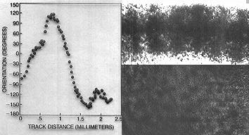
Hubel and Wiesel's original Nobel-winning work on occular processing columns
in the primary visual cortex showing millimetre-sized vertical conlumnar
organisation of line orientation detection in the anaesthesized cat. Left:
Changes in angle of optimal stimulation. Right Deoxyglucose radiographs
of vertical (above) and horizontal (below) sections. Scientific American.
Occular dominance columns were highlighted by injecting radio-actively labelled
amino-acids into the right eye of an anaesthetised animal. Scientific American.
The diversity of parallel visual processing
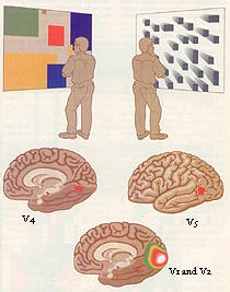
DISSIMILAR IMAGES stimulate different regions of the visual cortex. A brightly colored Mondrian causes area V4 to become highly active, as shown by tests of regional cerebral blood flow. Black-and-white moving images trigger activity in area V5. Both types of images lead to activity in areas VI and V2, which have less specialized functions and distribute signals to other cortical areas. S. Zeki. Scientific American.
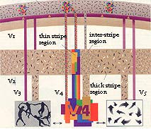
FOUR PERCEPTUAL PATHWAYS within the visual cortex have been identified.
Color is seen when wavelength-selective cells in the blob regions of VI
send signals to specialized area V4 and also to the thin stripes of V2,
which connect with V4. Form in association with color depends on connections
between the interblobs of VI, the interstripes of V2 and area V4. Cells
in layer 4B of Vl send signals to specialized areas V3 and V5 directly and
also through the thick stripes of V2; these connections give rise to the
perception of motion and dynamic form. Scientific American.
The World Seen through a Damaged Cortex
Damage to specialized regions of the cortex can cause strange types of blindness in which patients lose the ability to see just one attribute of the visual world, such as color, form or motion. Artwork produced by some of these patients offers glimpses into their view of the world, as well as into the workings of the visual cortex itself.

A patient with damage to the color pathways in the cortex lost all color vision. In his drawings, a banana, a tomato and green leaves all have similar colors. Scientific American.
This stroke patient suffered damage to the prestriate cortex that impaired his perception of form. He could copy a drawing with great skill, but he was unable to understand that the lines he had drawn produced an image of St. Paul's Cathedral. Scientific American.
Representation of the Surface of the Body in the Cortex
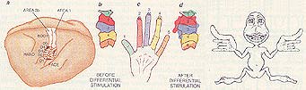
The homunculus ("little man") is a traditional way of illustrating how the surface of the body is represented in the somatosensory cortex. Larger areas of the cortex are devot ed to parts of the body that have greater sensitivity, such as the fingers and lips. Recently, the effects of sensitivity training have been shown in the owl monkey. The monkey's digits are represented in areas 3b and 1 of the somatosensory cortex (a). The diagrams (b and d) outline the regions that map the surface of the digits of an adult monkey (c) before and after training. During training the monkey rotated a disk for one hour a day, using only digits 2, 3 and occasionally 4. After three months of this activity, the area representing the stimulated fingers in the brain had increased substantially.
Working memory and the prefrontal cortex

NEURONAL CIRCUITRY connects the prefrontal cortex to the sensory, limbic and motor systems in a monkey brain (top). Anatomic studies show that neural projections from the parietal lobe to the prefrontal cortex exhibit a modular pattem, as seen in this frontal cross section (middle). Radioactive tracers reveal the metabolic activity in a frontal cross section of the brain of a monkey performing a delayed-response task (bottom). The distribution of activity closely conforms to the anatomic links. Scientific American.
Anomalies in the pre-frontal area may not result in noticeable differences to conventional intelligence scores, but they can profoundly disrupt a person's social capacity to plan and organise their life effectively, resulting in loss of employment and family life. They may become literally unable to make the simplest decision about choosing between an appointment on Tuesday andFriday because they cannot feel or express a preferance and have to cycle through labyrinths of logical reflection to no end because both choices are viable.
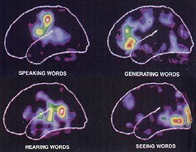
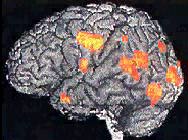
PET SCANS show the brain of a human subject performing a series of intellectual tasks related to words. The positron emission tomographic technique reveals that blood flow in the brain shifts to different locations, depending on which task is being performed. Scientific American.
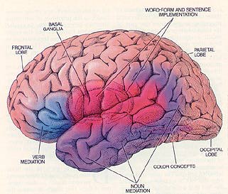
BRAIN SYSTEMS FOR LANGUAGE in the left hemisphere include word and sentence-implementation structures and mediation structures for various leidcal items and gramnw. The collections of neural structures that represent the concepts themselves are distributed across both right and left hemispheres in many sensory and motor regions. Scientific American.
A good idea of the plasticity ofthe brain, even in adults came from PET studies of a person who was fluent in both English and Spanish who then became a real-time live translator for the United Nations. After taking up this position, she developed completely new areas of her visual cortex so that she could think simultaneously in both languages.
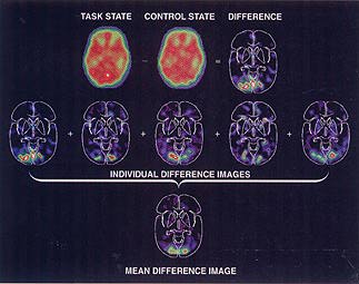
IMAGE SUBTRACTION AND AVERAGING serve as the foundation of functional brain imaging. Researchers subtract the PET blood-flow pattern of a control state from that of a task state to produce a difference image (top row). Data from different subjects are averaged (bottom two rows) to eliminate statistical fluctuations. Scientific American.
BLOOD FLOW to the brain provides the signals detected by functional MRI magnetic resonance imaging (nuclear magnetic resonance) and PET (positron-emission tomography). When resting neurons become active, blood flow to them increases. MRI detects changes in oxygen levels, which rise in the nearby blood vessels because active neurons consume no more oxygen than when they are at rest. PET relies on the increased delivery of injected radioactive water, which diffuses out of the vessels to reach all parts of the brain.
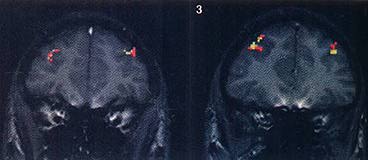
ACTIVE NEURAL AREAS from a subject remembering a sequence of letters are mapped by magnetic resonance imaging. The images below represent six slices through the frontal cortex. The slices are identified by numbers in the comers that correspond to those in the scan at the left. Red, orange and yellow represent areas of increasing activity. The resolution of MRI runs down to 1 mm, similar to the size of the cortical columnar organization detected by Hubel and Wiesel. Scientific American.
PET scan of cerebral excitation in a schizophrenic subject during hallucination.
